Lymphocytic Infiltration in the Heart Muscle, Thyroid Gland and Lung Following COVID-19 Injections
An excerpt from Taylor Hudak’s interview with Prof. Arne Burkhardt
Pathologist Prof. Dr. Arne Burkhardt discusses three findings in which he observed lymphocytic infiltration in the heart muscle, thyroid gland and lung.
Prof. Burkhardt observed myocarditis and other heart complications in his own study in which he examined the autopsy materials of patients who died shortly after vaccination.
In the first case, Prof. Burkhardt explains the observed changes to the heart muscle tissue in a patient with lymphocytic myocarditis. He compares the sample of heart muscle tissue to a normal sample.
Out of the 75 autopsies performed, Prof. Burkhardt had 31 cases in which the patient died from heart failure, including rhythmogenic failure. Of those 31 cases, 15 patients presented with perimyocarditis and 16 patients presented with a microangiopathy which is a disease of the small vessels unrelated to atherosclerosis.
“On the left side, you see the muscle cells which are elongated and have these long nuclei, but on the right you see in the middle between the muscle cells there are small blue dots which are the nuclei of lymphocytes. Lymphocytes are immunologically active cells and apparently they have been attracted by some antigenic material that is in the heart.”
Prof. Burkhardt explains that is it normal to observe one or two lymphocytes in a section of the heart muscle tissue. However, the presence of clusters of lymphocytes as observed in the image on the right is abnormal.
He compares the presence of lymphocytes in a sample to police officers patrolling a city.
“If you see one policeman or policewoman, that’s ok. That’s normal. If you see two, that’s still not alarming. But if you all of a sudden see 50 policemen, then you know there must be some trouble somewhere in the city.”
This same concept can be applied to understand the presence of lymphocytes in the heart muscle. If Prof. Burkhardt sees one or two lymphocytes, he says this is normal. But if the lymphocytes are aggregated in clumps as observed in the image on the right, this is indicative of inflammation, and in this case, myocarditis.
During his 40 years as a pathologist, Prof. Burkhardt observed one or two cases of myocarditis out of 1,500 to 2,000 autopsies performed yearly.
Lymphocytic infiltration of lung tissue
Clusters of lymphocytes were also observed in the lung tissue of an 82 year-old woman who died 40 days after receiving a second Moderna injection.
“You can see a small vessel and there’s lymphocytic infiltration around it which is not normally in the lung. And so this person definitely must have had some deficit in the gas exchange of the lung.”
Lymphocytic destruction of thyroid gland
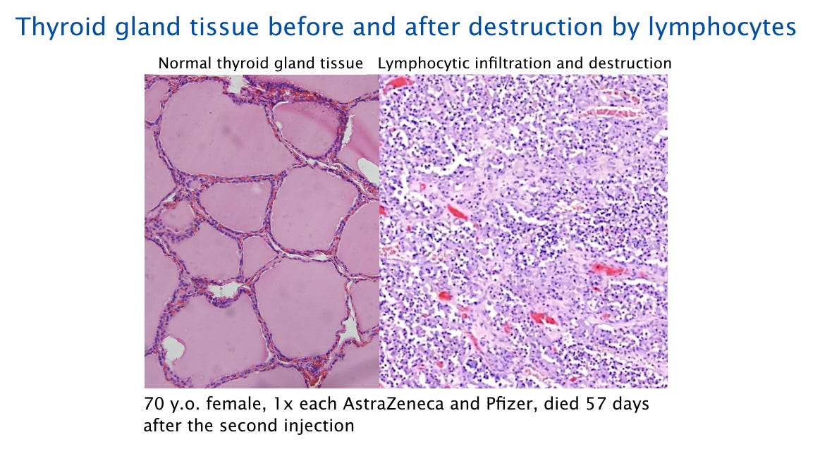
Striking changes to the thyroid gland tissue was observed in a 70 year-old woman who died 57 days after receiving a second COVID-19 injection.
“On the left side you see what we call follicles, which contain the thyroid hormone and the hormone is of course needed for the body. And on the right side, you see that these structures are lacking and instead of these structures there’s a lymphocytic infiltration which we have already seen in the other pictures. The lymphocytes destroy the thyroid tissue.”
This is similar to a well-known thyroid disease, Hashimoto’s Thyroiditis, which can occur without vaccination. However, Prof. Burkhardt says he is observing this condition more often since the introduction of the mRNA injections. Furthermore, in this case, the woman did not have thyroid disease and did not have a pre-existing condition that could have put her at risk for developing one.
Prof. Burkhardt says that damage to the thyroid gland tissue is irreversible. However, this condition can be treated through hormone medication.
How do mRNA vaccines cause lymphocytes to attack healthy cells?
The lymphocytes, a type of white blood cell, are a common theme among these three findings and will continue to be discussed in the subsequent cases.
An mRNA vaccine particle consists of a modified RNA molecule, which is contained in an envelope of fat-like molecules or lipids.
Once the vaccine has been injected and comes into contact with the body cells, the lipids which encase the mRNA molecule help the mRNA traverse the membrane which surrounds the cell allowing it to enter the cell.
The mRNA binds to the ribosomes within the cell, which are the cells’ little protein factories. The ribosomes read the information on the mRNA and create multiple copies of the spike protein molecule.
Intact spike protein molecules will transport to the cell surface, whereas some spike protein molecules are fragmented, and the fragments are taken to the cell surface.
There they are presented to the cells of the immune system by a specific carrier molecule, called MHC1. Think of MHC1 as a passport and the antigenic peptide, or the spike protein fragment carried by MHC1, as the individual details printed within the passport such as name and photograph.
T-lymphocytes which happen to possess T-cell receptors which match these antigenic peptides, or spike protein fragments, will recognize the MHC1 in combination with the spike protein fragments it carries, and then bind to it.
If a cytotoxic T-cell recognizes and binds its matching antigenic peptide, then it will attack and destroy the cell which presents it. This is a necessary step in antiviral defense. However, in the context of vaccination, it is unnecessary and potentially dangerous, as the immune system will attack healthy cells.
Lymphocytes occur in the spleen and the lymph nodes but are also in the blood. As we have seen before, the lymphocytes are fairly small, are round, and are typically stained dark purple. If lymphocytes appear in large quantities in tissues other than the lymph nodes or the spleen, this usually means that either a viral infection or some autoimmune disease is in progress.
A third possibility would be the rejection of a transplanted organ. Now we must contemplate another, novel mechanism, namely, the attack of the immune system on the vaccine expressing cells.
Destruction of cells that express the spike protein will also release cellular protein molecules which are normally hidden from the immune system. This will promote autoimmune diseases such as Hashimoto's thyroiditis.


