Rapid Aging, Scarring of the Arteries: A Side Effect of the COVID Injections
An excerpt from Taylor Hudak’s interview with Prof. Arne Burkhardt.
“Now my fear is that somebody, who may have a scar in his artery, maybe will die in five years from cerebral bleeding, but nobody will associate this with the vaccination…” - Prof. Arne Burkhardt
Watch Taylor Hudak’s full interview with Prof. Burkhardt here.
Written by Taylor Hudak
The elastic fibers, which consist of the protein elastin, are present in the skin, lungs, veins, arteries and other tissues in the body. These structures are formed in the first few years of life and very few to no elastic fibers are formed after puberty. Elastic fibers are permanent structures and are necessary for proper lung and heart function. However, once they are destroyed, they do not regenerate in adults.
Destruction of the elastic fibers can lead to several complications including emphysema in the lung. Additionally, loss of vascular elasticity may lead to hypertension or high blood pressure.
“It’s very important for the arteries, especially the main artery, the aorta, because it gives elasticity,” said Prof. Burkhardt. “It’s important in the lung because it gives elasticity to help with breathing.”
Damage to the Aorta
The walls of the aorta and of other major arteries are rich in elastic fibers which are arranged into stacked layers or lamellae. The elastic lamellae are essential for the vessels ability to withstand the pulsating blood pressure. Prof. Burkhardt found that in many of his cases, the elastic lamella were damaged and disrupted particularly within the hotspots of inflammation.
“If the arteries were not elastic, we would have peaks in the blood pressure, and these peaks, of course, may lead to rupture.”
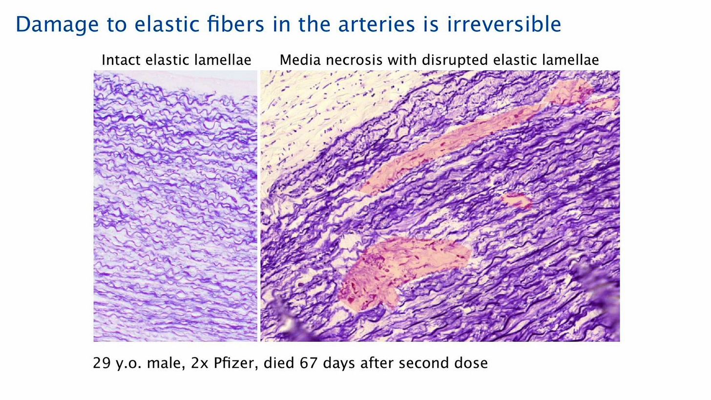
Damage to the aorta and other major arteries was also apparent in patients who had not suffered overt failure or rupture to these vessels. The image below shows the aortic wall of a 29 year-old male who died 67 days after the second Pfizer injection. The tissue sample had been treated with a special stain which highlights the disrupted elastic lamellae. The image on the left shows intact (healthy) elastic lamellae for a comparison.

The image on the left shows the intact elastic lamellae of a normal healthy artery, which is constructed of a very regular stratification (layering) of myofibroblasts (cells that form and maintain connective tissue), smooth muscle cells and the elastic fibers.
“We have very alarming findings — first of all, destruction of elastic fibers in the arteries — especially in the aorta.”
The image on the right shows very small lesions, which are highlighted in pink. Patients with these lesions do not not always have symptoms. However, those that have a further development of the lesions — that become a total media necrosis of the elastic fibers — are at risk of a fatal aortic rupture.
Prof. Burkhardt observed five aortic ruptures in this series of 75 autopsies in vaccinated patients.
Anatomy of an Artery
The larger arteries, including the aorta, consist of three layers. The innermost layer is the intima, which is the layer most often affected by arteriosclerosis and cholesterol deposition. The media, the middle layer, is where the elastic fibers, the myofibroblasts and the smooth muscle cells are located. The adventitia, the outermost layer, contains the vasa vasorum, which are small vessels that supply the wall of the blood vessel itself with blood.
This concerns in particular the outer layers of the blood vessel walls; in contrast, the inner layers are supplied mainly by diffusion from the blood that flows through the lumen of the vessel itself. The middle layer — the deep media — is the furthest removed from the supply in either direction. This makes it particularly vulnerable to toxic agents, including infectious toxic agents. One possible outcome is infectious toxic media necrosis, a weakening of the vessel wall which may lead to rupture.
Mechanism of Harm
100 years ago, infectious toxic media necrosis was often seen in patients with syphilis. This condition put the infected person at risk for a potentially fatal rupture of the artery.
Lathyrism, which is a toxic damage to the blood vessels and sometimes bones and the nervous system due to excessive consumption of chickpeas, is a type of food poisoning that may lead to toxic media necrosis. Prof. Burkhardt said that during his first years as a pathologist he observed lathyrism in some patients, but that it is rarely seen today.
According to Prof. Burkhardt, the same mechanism of infectious toxic media necrosis that was observed in the past, is likely similar to what is being observed today in vaccinated patients who develop media necrosis. This also involves the destruction of the elastic fibers, which are crucial for the function and the stability of the aorta.
“There is a toxic and maybe also immunologic attack in the area of the arteries where there is a weak point, and there may be local bleeding with Hemosiderosis, (excessive accumulation of iron deposits in the tissue) and there may be perforation.”
However, in some cases, the small lesions may heal. But in other cases, in which the elastic fibers cannot be replaced (once the patient is an adult), it can leave a scar. If there is a scar, the artery loses its elasticity and so the rise in the blood pressure during the contraction of the heart is very high and it goes up and down repeatedly. Given that the brain arteries are the most sensitive, it may lead to rupture and cause death by cerebral bleeding.
“My fear is that someone who has this scar in his (brain) artery, maybe he will die in five years from cerebral bleeding, but nobody will associate this with the vaccination, and no one will even examine the aorta. It is not a standard to examine the aorta.”
Prof. Burkhardt suggests that there may be a high number of cases in which no one will see a connection with the vaccination although it is probable.
Advanced Aging
Additionally, the elastic fibers are present in the skin. As one gets older, the elastic fibers become damaged through exposure to ultra violet radiation, which contributes to the physical appearance of aging.
At the time of filming the interview, Prof. Burkhardt was systematically reviewing biopsies of the skin in which he observed damage to the elastic fibers in vaccinated persons.
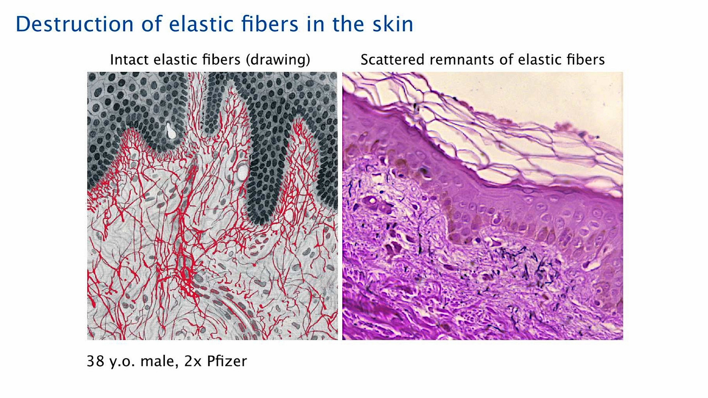
The right-hand image below shows the destruction of elastic fibers in the skin of a 38 year-old man, who received two Pfizer injections. The image on the left shows intact elastic fibers for a comparison.
“You see, on the left side, there’s a very delicate network of very fine elastic fibers (highlighted in red), and on the top, is the epithelium of the dermis.”
The patient, whose skin biopsy is shown on the right, has vasculitis of the skin. The very delicate black lines (see video) are the remnants of the elastic fibers. Additionally, there is no network below the basement membrane.
“There have been very convincing reports that people after the vaccination suddenly appear to look much older,” said Prof. Burkhardt. “Now this may be due to psychological factors too, but we definitely have proof that these elastic fibers in some cases are profoundly destroyed in the skin.”


Love Taylor from the days of Julian's kidnapping from the Embassy & she should remember me from pre-2020 Twitter ban that endures but Biotech Mafia is my long suit and this is not the source she needs to get to the truth.. ping me and skip the acadamagicians entirely! :~)